
Quantitative Prediction of Spirulina platensis Biomass Using UV-Vis Spectrophotometry
DOI:
https://doi.org/10.55981/limnotek.2026.14459Keywords:
Spirulina platensis, Spectrophotometer, Optical DensityAbstract
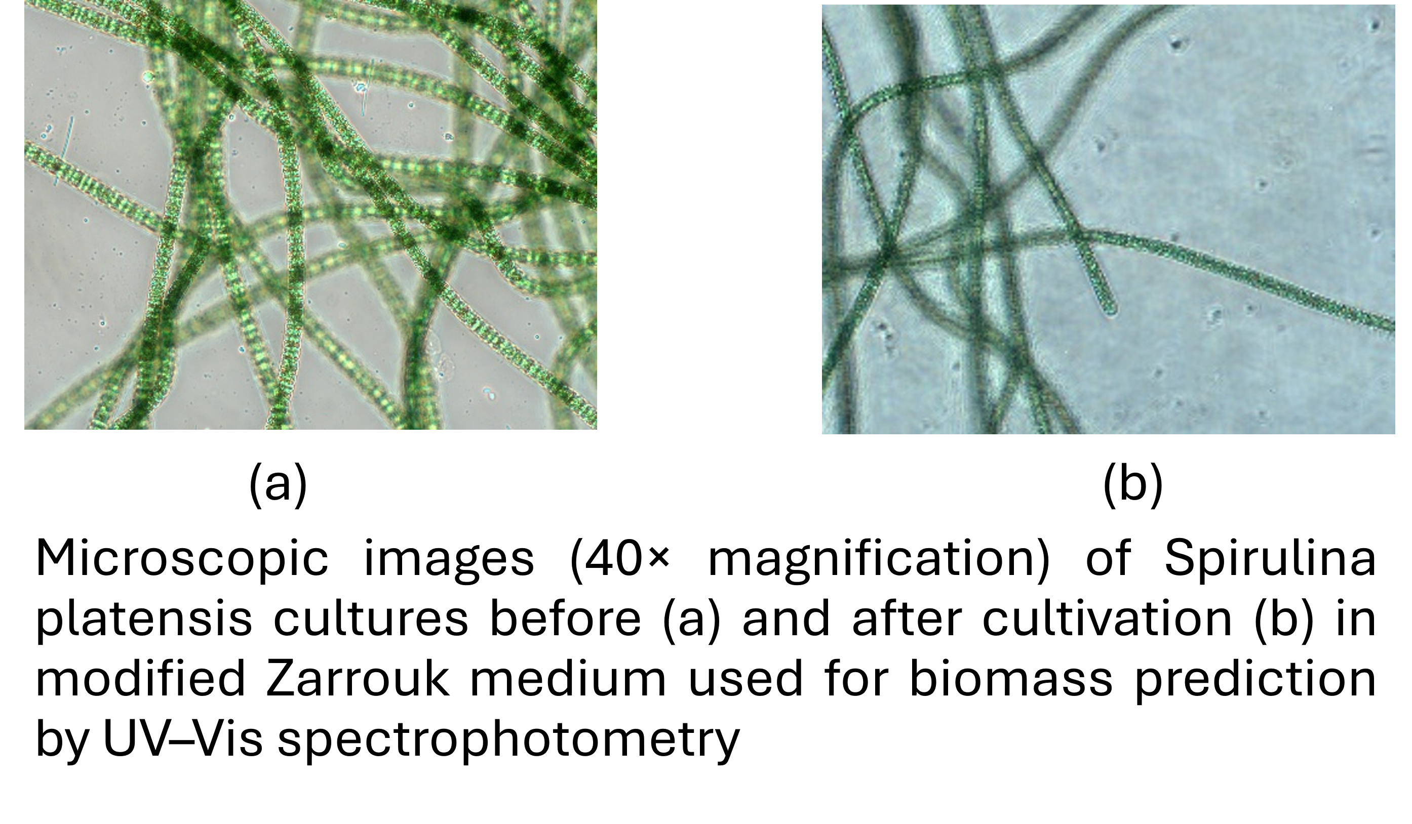
Spirulina platensis has become a promising feedstock for the synthesis of several industrially important biomolecules, including proteins, lipids, and carotenoids. However, significant technological obstacles pertaining to optimization, growth monitoring, and biomolecule extraction in Spirulina remain despite advancements in industrial-scale microalgae production and biomolecule harvesting. Standard techniques used for microalgal biomass and biomolecules monitoring include FTIR spectroscopy, colorimetric techniques, and manual cell counting. However, these techniques have drawbacks, particularly processing time and handling errors. This paper seeks to establish an operational equation that effectively relates measured absorbance (or optical density, OD) to the dry weight of Spirulina platensis microalgae using a UV-Vis spectrophotometer. The wavelengths of 680, 750, and 565 nm were selected based on the absorption spectrum of chlorophyll-a, as well as the wavelengths at which absorbance does not reach its peak. The best results were obtained at a wavelength of 680 nm with the equation y = 1.2759x + 0.1512, with an R² value of 0.9914. This technique allows for more accurate measurement of Spirulina platensis dry weight and total biomass.
References
Agberien, A. V., & Örmeci, B. 2020. Monitoring of Cyanobacteria in Water Using Spectrophotometry and First Derivative of Absorbance.
AlFadhly, N. K. Z., Alhelfi, N., Altemimi, A. B., Verma, D. K., & Cacciola, F. 2022. Tendencies affecting the growth and cultivation of genus Spirulina: an investigative review on current trends. Plants, 11(22), 1–21. https://doi.org/10.3390/plants11223063
Aparicio, S., Pach, M., & Borr, L. 2022. Comprehensive assessment of the microalgae-nitrifying bacteria competition in microalgae-based wastewater treatment systems : Relevant factors , evaluation methods and control strategies. 61(June 2021). https://doi.org/10.1016/j.algal.2021.102563
APHAAWWA. 2017. Standard Methods. In L. S. C. Arnold E. Greenberg, R. Rhodes Trussell (Ed.), Standar methods for the examination of water and wastewater (23rd ed.). Washington, DC. https://doi.org/10.1016/B978-0-12-382165-2.00237-3
Barth, D., Maia, E., Hyttinen, E., & Timo, D. 2025. Spectrochimica Acta Part A : Molecular and Biomolecular Spectroscopy Biological contaminants analysis in microalgae culture by UV – vis spectroscopy and machine learning. 330(September 2024). https://doi.org/10.1016/j.saa.2024.125690
Bartošová, A., & Blinová, L. 2015. Faculty Of Materials Science And Technology In Trnava CHARACTERISATION OF POLYSACHARIDES AND LIPIDS FROM SELECTED GREEN ALGAE SPECIES BY FTIR-ATR. 23(36), 97–102.
Baweja, P., & Sahoo, D. 2015. No Title. In D. Sahoo & J. Seckbach (Eds.), the algae world. Cellular Origin, Life in Extreme Habitats and Astrobiology. Springer. https://doi.org/10.1007/978-94-017-7321-8_2
Caprio, F. Di. 2020. Methods to quantify biological contaminants in microalgae cultures. Algal Research, 49(May), 101943. https://doi.org/10.1016/j.algal.2020.101943
Chrismadha, T., Satya, A., Satya, I. A., Rosidah, R., Satya, A. D. M., Pangestuti, R., Harimawan, A., Setiadi, T., Chew, K. W., & Show, P. L. 2022. Outdoor Inclined Plastic Column Photobioreactor: Growth, and Biochemicals Response of Arthrospira platensis Culture on Daily Solar Irradiance in a Tropical Place. Metabolites, 12(12), 1–14. https://doi.org/10.3390/metabo12121199
Dziosa, K., & Makowska, M. 2016. MONITORING OF CHLORELLA sp . GROWTH BASED. Problemy Eksploatacji, 2, 197–206.
Eddiwan, K., Dahril, T., & Efawani, E. 2023. Relationship Between Pigment Concentration and Dry Weight in Determining Microalgae Abundance in Artificial Water Samples. International Journal of Research and Scientific Innovation, X(V), 118–127. https://doi.org/10.51244/ijrsi.2023.10512
Elisabeth, B., Rayen, F., & Behnam, T. 2021. Critical Reviews in Biotechnology Microalgae culture quality indicators : a review. Critical Reviews in Biotechnology, 41(4), 457–473. https://doi.org/10.1080/07388551.2020.1854672
Fakhri, M., Riyani, E., Ekawati, A. W., Arifin, N. B., Yuniarti, A., Widyawati, Y., Saputra, I. K., Samuel, P. D., Arif, M. Z., & Hariati, A. M. 2021. Biomass, pigment production, and nutrient uptake of chlorella sp. Under different photoperiods. Biodiversitas, 22(12), 5344–5349. https://doi.org/10.13057/biodiv/d221215
Ferreira, S., & Sant, C. 2017. Impact of culture conditions on the chlorophyll content of microalgae for biotechnological applications. World Journal of Microbiology and Biotechnology. https://doi.org/10.1007/s11274-016-2181-6
Goher, M. E., El-monem, A. M. A., Abdel-satar, A. M., Ali, M. H., Hussian, A. M., & Napiórkowska-krzebietke, A. 2016. BIOSORPTION OF SOME TOXIC METALS FROM AQUEOUS SOLUTION USING NON-LIVING ALGAL CELLS. https://doi.org/10.5601/jelem.2015.20.4.1037
Grif, M. J., Garcin, C., Hille, R. P. Van, & Harrison, S. T. L. 2011. Interference by pigment in the estimation of microalgal biomass concentration by optical density. 85, 119–123. https://doi.org/10.1016/j.mimet.2011.02.005
Guiry, M.D. & Guiry, G.M. 2026. AlgaeBase. World-wide electronic publication, University of Galway. https://www.algaebase.org; Accesed on 5 February, 2026
Heriberto, J., Zuluaga, G., Salazar, A., Diez, G., Gomez, A., Martínez, T., & Mariana, J. F. V. 2018. Automatic identification of Scenedesmus polymorphic microalgae from microscopic images. Pattern Analysis and Applications, 21(2), 601–612. https://doi.org/10.1007/s10044-017-0662-3
Hotos, G. N., Avramidou, D., & Bekiari, V. 2020. Calibration Curves of Culture Density Assessed by Spectrophotometer for Three Microalgae (Nephroselmis sp., Amphidinium carterae and Phormidium sp.). European Journal of Biology and Biotechnology, 1(6), 1–7. https://doi.org/10.24018/ejbio.2020.1.6.132
Katam, K., Ananthula, R., Anumala, S., Sriariyanun, M., & Bhattacharyya, D. 2022. The impact of light intensity and wavelength on the performance of algal-bacterial culture treating domestic wastewater. 02003.
Liu, J., Zeng, L., & Ren, Z. 2019. Recent application of spectroscopy for the detection of microalgae life information : A review. Applied Spectroscopy Reviews, 0(0), 1–34. https://doi.org/10.1080/05704928.2018.1509345
Mahlangu, D., Mphahlele, K., & Paola, F. De. 2024. Microalgae-Mediated Biosorption for Effective Heavy Metals Removal from Wastewater : A Review. 1–23.
Malhotra, A., & Ormeci, B. 2023. Journal of Photochemistry & Photobiology , B : Biology Detection and identification of a mixed cyanobacteria and microalgae culture using derivative spectrophotometry. 238(November 2022). https://doi.org/10.1016/j.jphotobiol.2022.112616
Malletzidou, L., Kyratzopoulou, E., Kyzaki, N., Nerantzis, E., & Kazakis, N. A. 2024. Near-Infrared Spectroscopy for Growth Estimation of Spirulina platensis Cultures.
Mori, A., Yamashita, K., Tabata, Y., Seto, K., & Tokunaga, E. 2021. Absorbance spectroscopy of light scattering samples placed inside an integrating sphere for wide dynamic range absorbance measurement Absorbance spectroscopy of light scattering samples placed inside an integrating sphere for wide dynamic range absorbance measurement. 123103. https://doi.org/10.1063/5.0066412
Myers, J. A., Curtis, B. S., & Curtis, W. R. 2013. Improving accuracy of cell and chromophore concentration measurements using optical density Improving accuracy of cell and chromophore concentration measurements using optical density.
Nagabhushan, C. M. 2023. UV-Spectroscopic Studies on the Potential State of Spirulina Cultures using Varied Culture Media in Uncontrolled Laboratory Conditions in Ballari Region. 3(2), 70–76. https://doi.org/10.48175/IJARSCT-7953
Nanni, M. R. (2023). A Novel Method for Estimating Chlorophyll and Carotenoid Concentrations in Leaves : A Two Hyperspectral Sensor Approach.
Nielsen, S. L., & Hansen, B. W. 2019. Evaluation of the robustness of optical density as a tool for estimation of biomass in microalgal cultivation : The effects of growth conditions and physiological state. April, 2698–2706. https://doi.org/10.1111/are.14227
Nyakundi, D. O., & Cleophas, P. 2021. Harnessing nutritional benefits of Spirulina platensis: standardization of cultivating conditions of Spirulina in Kilimanjaro. Tanzania Journal of Science, 47(4), 1412–1423. https://doi.org/10.4314/tjs.v47i4.7
Pahija, E., & Hui, C. 2019. A systematic study on the effects of dynamic environments on microalgae concentration. Algal Research, 42(April), 101599. https://doi.org/10.1016/j.algal.2019.101599
Phansi, P., Ferreira, S., & Cerd, V. 2022. Trends in Analytical Chemistry From mono- to multicomponent methods in UV-VIS spectrophotometric and fl uorimetric quantitative analysis e A review. 157. https://doi.org/10.1016/j.trac.2022.116772
Podevin, M., Fotidis, I. A., Angelidaki, I., Podevin, M., & Fotidis, I. A. 2017. Critical Reviews in Biotechnology Microalgal process-monitoring based on high- selectivity spectroscopy tools : status and future perspectives status and future perspectives. Critical Reviews in Biotechnology, 0(0), 1–15. https://doi.org/10.1080/07388551.2017.1398132
Richmond, A., & Hu, Q. (Eds.). 2013. handbook of microlagal culture. John Wiley & Sons.
Rinanti, A., Fachrul, M. F., Hadisoebroto, R., Minarti, A., & Sunaryo, T. 2022. Increasing Effectiveness of Heavy Metal Sorption by Biosorbent Microalgae Beads. 23(7), 50–57.
Salgueiro, J. L., Pérez, L., Maceiras, R., Sánchez, Á., & Cancela, Á. 2018. Semicontinuous culture of Chlorella vulgaris microalgae for wastewater treatment. International Journal of Environmental Research, 12, 765–772. https://doi.org/10.1007/s41742-018-0129-4
Santos-Ballardo, D. U., Rossi, S., Hernández, V., Gómez, R. V., del Carmen Rendón-Unceta, M., Caro-Corrales, J., & Valdez-Ortiz, A. 2015. A simple spectrophotometric method for biomass measurement of important microalgae species in aquaculture. Aquaculture, 448, 87–92. https://doi.org/10.1016/j.aquaculture.2015.05.044
Schagerl, M., Siedler, R., Konop, E., & Ali, S. S. 2022. Estimating Biomass and Vitality of Microalgae for Monitoring Cultures : A Roadmap for Reliable Measurements.
Soni, R. A., Sudhakar, K., & Rana, R. S. 2017. Spirulina – From growth to nutritional product: A review. Trends in Food Science and Technology, 69, 157–171. https://doi.org/10.1016/j.tifs.2017.09.010
Sudhakar, K., & Premalatha, M. 2015. Environmental Effects Characterization of Micro Algal Biomass Through FTIR / TGA / CHN Analysis : Application to Scenedesmus sp . TGA / CHN Analysis : Application to Scenedesmus sp . 7036(December). https://doi.org/10.1080/15567036.2013.825661
Taleuzzaman, M. 2018. Limit of Blank ( LOB ), Limit of Detection ( LOD ), and Limit of Quantification ( LOQ ). 7(5). https://doi.org/10.19080/OMCIJ.2018.07.555722
Tavˇ, P., & Dolinar, M. 2025. A Pipeline for the Isolation and Cultivation of Microalgae and Cyanobacteria from Hypersaline Environments. 13–15.
Tiwari, S., Sharma, V., Patel, A., Yadav, V., Kalra, C., Singh, G., Arif, N., Khandelwal, A., Tiwari, A., & Singh, D. K. 2026. Microalgae : A valuable bio-resource for pharmaceuticals and nutraceuticals. 14(1), 5–17. https://doi.org/10.7324/JABB.2025.258624
Wacogne, B., Podevin, M. B., Vaccari, N., Koubevi, C., Gutierrez, E., Davoine, L., Robert-nicoud, M., Rouleau, A., & Frelet-barrand, A. 2024. Concentration vs . Optical Density of ESKAPEE Bacteria : A Method to Determine the Optimum Measurement Wavelength.
Yap, P. Y., Jain, A., & Trau, D. 2018. an 050 Life Sciences Determination of Biomass in Spirulina Cultures By Photopette . February, 1–3. www.tipbiosystems.com
Downloads
Published
Issue
Section
License
Copyright (c) 2026 Fajar Sumi Lestari, Agustino Zulys, Awalina Satya

This work is licensed under a Creative Commons Attribution 4.0 International License.